Sitemap

A list of all the posts and pages found on the site. For you robots out there is an XML version available for digesting as well.

Pages

Posts

Future Blog Post

This post will show up by default. To disable scheduling of future posts, edit config.yml and set future: false.

Blog Post number 4

This is a sample blog post. Lorem ipsum I can’t remember the rest of lorem ipsum and don’t have an internet connection right now. Testing testing testing this blog post. Blog posts are cool.

Blog Post number 3

This is a sample blog post. Lorem ipsum I can’t remember the rest of lorem ipsum and don’t have an internet connection right now. Testing testing testing this blog post. Blog posts are cool.

Blog Post number 2

This is a sample blog post. Lorem ipsum I can’t remember the rest of lorem ipsum and don’t have an internet connection right now. Testing testing testing this blog post. Blog posts are cool.

Blog Post number 1

This is a sample blog post. Lorem ipsum I can’t remember the rest of lorem ipsum and don’t have an internet connection right now. Testing testing testing this blog post. Blog posts are cool.

news

Welcome to our new members

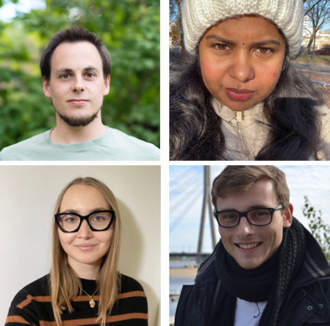
We welcome our new team members:
Dr. Alessandro Enrico is a materials scientist from the KTH Royal Institute of Technology in Stockholm (Sweden),
Dr. Saranya Vasudevan is an expert in molecular dynamics from Bharathiar University (India),
Ms. Bohdana Horda from Unipv (Italy) is a graduate of our MS in Bioengineering at the University of Pavia (Italy),
Dr. Julius Zimmermann is a physicist and computational scientist from the University of Rostock (Germany).

New grants

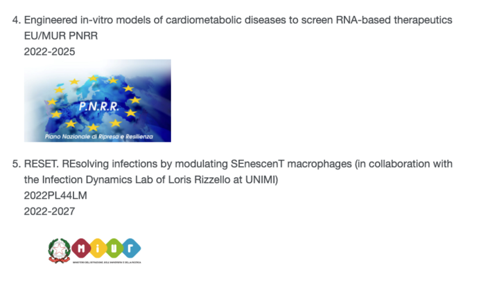
The lab was fortunate to secure new funding for collaborative projects:

  • Together with Prof. Priori at UNIPV/CNIC, we will develop engineered heart-chips for testing RNA-therapeutics in the context of the EU/PNRR-funded National Center for Gene Therapy and RNA-based medicine.
  • Together with Prof. Rizzello at UNIMI, we will develop a tuberculosis on-a-chip model to study the mechanobiology of senescent macrophages.

publications

Matrix elasticity regulates the optimal cardiac myocyte shape for contractility

Abstract

Concentric hypertrophy is characterized by ventricular wall thickening, fibrosis, and decreased myocyte length-to-width aspect ratio. Ventricular thickening is considered compensatory because it reduces wall stress, but the functional consequences of cell shape remodeling in this pathological setting are unknown. We hypothesized that decreases in myocyte aspect ratio allow myocytes to maximize contractility when the extracellular matrix becomes stiffer due to conditions such as fibrosis. To test this, we engineered neonatal rat ventricular myocytes into rectangles mimicking the 2-D profiles of healthy and hypertrophied myocytes on hydrogels with moderate (13 kPa) and high (90 kPa) elastic moduli. Actin alignment was unaffected by matrix elasticity, but sarcomere content was typically higher on stiff gels. Microtubule polymerization was higher on stiff gels, implying increased intracellular elastic modulus. On moderate gels, myocytes with moderate aspect ratios (∼7:1) generated the most peak systolic work compared with other cell shapes. However, on stiffer gels, low aspect ratios (∼2:1) generated the most peak systolic work. To compare the relative contributions of intracellular vs. extracellular elasticity to contractility, we developed an analytical model and used our experimental data to fit unknown parameters. Our model predicted that matrix elasticity dominates over intracellular elasticity, suggesting that the extracellular matrix may potentially be a more effective therapeutic target than microtubules. Our data and model suggest that myocytes with lower aspect ratios have a functional advantage when the elasticity of the extracellular matrix decreases due to conditions such as fibrosis, highlighting the role of the extracellular matrix in cardiac disease.

Download here

Origin of Cardiomyocytes in the Adult Heart

Abstract

This review article discusses the mechanisms of cardiomyogenesis in the adult heart. They include the re-entry of cardiomyocytes into the cell cycle; dedifferentiation of pre-existing cardiomyocytes, which assume an immature replicating cell phenotype; transdifferentiation of hematopoietic stem cells into cardiomyocytes; and cardiomyocytes derived from activation and lineage specification of resident cardiac stem cells. The recognition of the origin of cardiomyocytes is of critical importance for the development of strategies capable of enhancing the growth response of the myocardium; in fact, cell therapy for the decompensated heart has to be based on the acquisition of this fundamental biological knowledge.

Download here

Late Na+ current and protracted electrical recovery are critical determinants of the aging myopathy

Abstract

The aging myopathy manifests itself with diastolic dysfunction and preserved ejection fraction. We raised the possibility that, in a mouse model of physiological aging, defects in electromechanical properties of cardiomyocytes are important determinants of the diastolic characteristics of the myocardium, independently from changes in structural composition of the muscle and collagen framework. Here we show that an increase in the late Na+ current (INaL) in aging cardiomyocytes prolongs the action potential (AP) and influences temporal kinetics of Ca2+ cycling and contractility. These alterations increase force development and passive tension. Inhibition of INaL shortens the AP and corrects dynamics of Ca2+ transient, cell contraction and relaxation. Similarly, repolarization and diastolic tension of the senescent myocardium are partly restored. Thus, INaL offers inotropic support, but negatively interferes with cellular and ventricular compliance, providing a new perspective of the biology of myocardial aging and the aetiology of the defective cardiac performance in the elderly.

Download here

Mechanotransduction and Metabolism in Cardiomyocyte Microdomains

Abstract

Efficient contractions of the left ventricle are ensured by the continuous transfer of adenosine triphosphate (ATP) from energy production sites, the mitochondria, to energy utilization sites, such as ionic pumps and the force-generating sarcomeres. To minimize the impact of intracellular ATP trafficking, sarcomeres and mitochondria are closely packed together and in proximity with other ultrastructures involved in excitation-contraction coupling, such as t-tubules and sarcoplasmic reticulum junctions. This complex microdomain has been referred to as the intracellular energetic unit. Here, we review the literature in support of the notion that cardiac homeostasis and disease are emergent properties of the hierarchical organization of these units. Specifically, we will focus on pathological alterations of this microdomain that result in cardiac diseases through energy imbalance and posttranslational modifications of the cytoskeletal proteins involved in mechanosensing and transduction.

Download here

Heart Valve Replacements with Regenerative Capacity

Abstract

The incidence of severe valvular dysfunctions (e.g., stenosis and insufficiency) is increasing, leading to over 300,000 valves implanted worldwide yearly. Clinically used heart valve replacements lack the capacity to grow, inherently requiring repetitive and high-risk surgical interventions during childhood. The aim of this review is to present how different tissue engineering strategies can overcome these limitations, providing innovative valve replacements that proved to be able to integrate and remodel in pre-clinical experiments and to have promising results in clinical studies. Upon description of the different types of heart valve tissue engineering (e.g., in vitro, in situ, in vivo, and the pre-seeding approach) we focus on the clinical translation of this technology. In particular, we will deepen the many technical, clinical, and regulatory aspects that need to be solved to endure the clinical adaptation and the commercialization of these promising regenerative valves.

Download here

Coupling primary and stem cell–derived cardiomyocytes in an in vitro model of cardiac cell therapy

Abstract

The efficacy of cardiac cell therapy depends on the integration of existing and newly formed cardiomyocytes. Here, we developed a minimal in vitro model of this interface by engineering two cell microtissues (μtissues) containing mouse cardiomyocytes, representing spared myocardium after injury, and cardiomyocytes generated from embryonic and induced pluripotent stem cells, to model newly formed cells. We demonstrated that weaker stem cell–derived myocytes coupled with stronger myocytes to support synchronous contraction, but this arrangement required focal adhesion-like structures near the cell–cell junction that degrade force transmission between cells. Moreover, we developed a computational model of μtissue mechanics to demonstrate that a reduction in isometric tension is sufficient to impair force transmission across the cell–cell boundary. Together, our in vitro and in silico results suggest that mechanotransductive mechanisms may contribute to the modest functional benefits observed in cell-therapy studies by regulating the amount of contractile force effectively transmitted at the junction between newly formed and spared myocytes.

Download here

Phototactic guidance of a tissue-engineered soft-robotic ray

Abstract

Swim into the light
      <jats:p>
        A bio-inspired swimming robot that mimics a ray fish can be guided by light. Park
        <jats:italic>et al.</jats:italic>
        built a 1/10th-scale version of a ray fish with a microfabricated gold skeleton and a rubber body powered by rat heart muscle cells. The cardiomyocytes were genetically engineered to respond to light cues, so that the undulatory movements propelling the robot through water would follow a light source.
      </jats:p>
      <jats:p>
        <jats:italic>Science</jats:italic>
        , this issue p.
        <jats:related-article xmlns:xlink="http://www.w3.org/1999/xlink" ext-link-type="doi" issue="6295" page="158" related-article-type="in-this-issue" vol="353" xlink:href="10.1126/science.aaf4292">158</jats:related-article>
      </jats:p>

Download here

A human in vitro model of Duchenne muscular dystrophy muscle formation and contractility

Abstract

Tongue weakness, like all weakness in Duchenne muscular dystrophy (DMD), occurs as a result of contraction-induced muscle damage and deficient muscular repair. Although membrane fragility is known to potentiate injury in DMD, whether muscle stem cells are implicated in deficient muscular repair remains unclear. We hypothesized that DMD myoblasts are less sensitive to cues in the extracellular matrix designed to potentiate structure–function relationships of healthy muscle. To test this hypothesis, we drew inspiration from the tongue and engineered contractile human muscle tissues on thin films. On this platform, DMD myoblasts formed fewer and smaller myotubes and exhibited impaired polarization of the cell nucleus and contractile cytoskeleton when compared with healthy cells. These structural aberrations were reflected in their functional behavior, as engineered tongues from DMD myoblasts failed to achieve the same contractile strength as healthy tongue structures. These data suggest that dystrophic muscle may fail to organize with respect to extracellular cues necessary to potentiate adaptive growth and remodeling.

Download here

Organ Chips: Quality Assurance Systems in Regenerative Medicine

Abstract

A class of novel therapies leverages regenerative cell types in disease microenvironments. This complex interplay challenges established good manufacturing practices, as standards and analytical tools to measure regenerative potency are missing. That is, we can build the product right, but we do not know if we are building the right product. Here, we suggest that organ‐chips, biomimetic in vitro phenotyping platforms, can serve as key quality assurance systems in regenerative medicine.

Download here

Translational cardiac stem cell therapy: advancing from first-generation to next-generation cell types

Abstract

Acute myocardial infarction and chronic heart failure rank among the major causes of morbidity and mortality worldwide. Except for heart transplantation, current therapy options only treat the symptoms but do not cure the disease. Stem cell-based therapies represent a possible paradigm shift for cardiac repair. However, most of the first-generation approaches displayed heterogeneous clinical outcomes regarding efficacy. Stemming from the desire to closely match the target organ, second-generation cell types were introduced and rapidly moved from bench to bedside. Unfortunately, debates remain around the benefit of stem cell therapy, optimal trial design parameters, and the ideal cell type. Aiming at highlighting controversies, this article provides a critical overview of the translation of first-generation and second-generation cell types. It further emphasizes the importance of understanding the mechanisms of cardiac repair and the lessons learned from first-generation trials, in order to improve cell-based therapies and to potentially finally implement cell-free therapies.

Download here

Insights Into the Pathogenesis of Catecholaminergic Polymorphic Ventricular Tachycardia From Engineered Human Heart Tissue

Abstract

Background: Modeling of human arrhythmias with induced pluripotent stem cell–derived cardiomyocytes has focused on single-cell phenotypes. However, arrhythmias are the emergent properties of cells assembled into tissues, and the impact of inherited arrhythmia mutations on tissue-level properties of human heart tissue has not been reported.
      <jats:sec>
        <jats:title>Methods:</jats:title>
        <jats:p>Here, we report an optogenetically based, human engineered tissue model of catecholaminergic polymorphic ventricular tachycardia (CPVT), an inherited arrhythmia caused by mutation of the cardiac ryanodine channel and triggered by exercise. We developed a human induced pluripotent stem cell–derived cardiomyocyte–based platform to study the tissue-level properties of engineered human myocardium. We investigated pathogenic mechanisms in CPVT by combining this novel platform with genome editing.</jats:p>
      </jats:sec>
      <jats:sec>
        <jats:title>Results:</jats:title>
        <jats:p>
          In our model, CPVT tissues were vulnerable to developing reentrant rhythms when stimulated by rapid pacing and catecholamine, recapitulating hallmark features of the disease. These conditions elevated diastolic Ca
          <jats:sup>2+</jats:sup>
          levels and increased temporal and spatial dispersion of Ca
          <jats:sup>2+</jats:sup>
          wave speed, creating a vulnerable arrhythmia substrate. Using Cas9 genome editing, we pinpointed a single catecholamine-driven phosphorylation event, ryanodine receptor–serine 2814 phosphorylation by Ca
          <jats:sup>2</jats:sup>
          <jats:sup>+</jats:sup>
          /calmodulin-dependent protein kinase II, that is required to unmask the arrhythmic potential of CPVT tissues.
        </jats:p>
      </jats:sec>
      <jats:sec>
        <jats:title>Conclusions:</jats:title>
        <jats:p>Our study illuminates the molecular and cellular pathogenesis of CPVT and reveals a critical role of calmodulin-dependent protein kinase II–dependent reentry in the tissue-scale mechanism of this disease. We anticipate that this approach will be useful for modeling other inherited and acquired cardiac arrhythmias.</jats:p>
      </jats:sec>

Download here

A Pulsatile Flow System to Engineer Aneurysm and Atherosclerosis Mimetic Extracellular Matrix

Abstract

Alterations of blood flow patterns strongly correlate with arterial wall diseases such as atherosclerosis and aneurysm. Here, a simple, pumpless, close‐loop, easy‐to‐replicate, and miniaturized flow device is introduced to concurrently expose 3D engineered vascular smooth muscle tissues to high‐velocity pulsatile flow versus low‐velocity disturbed flow conditions. Two flow regimes are distinguished, one that promotes elastin and impairs collagen I assembly, while the other impairs elastin and promotes collagen assembly. This latter extracellular matrix (ECM) composition shares characteristics with aneurysmal or atherosclerotic tissue phenotypes, thus recapitulating crucial hallmarks of flow‐induced tissue morphogenesis in vessel walls. It is shown that the mRNA levels of ECM of collagens and elastin are not affected by the differential flow conditions. Instead, the differential gene expression of matrix metalloproteinase (MMP) and their inhibitors (TIMPs) is flow‐dependent, and thus drives the alterations in ECM composition. In further support, treatment with doxycycline, an MMP inhibitor and a clinically used drug to treat vascular diseases, halts the effect of low‐velocity flow on the ECM remodeling. This illustrates how the platform can be exploited for drug efficacy studies by providing crucial mechanistic insights into how different therapeutic interventions may affect tissue growth and ECM assembly.

Download here

SEM2: Introducing mechanics in cell and tissue modeling using coarse-grained homogeneous particle dynamics

Abstract

Modeling multiscale mechanics in shape-shifting engineered tissues, such as organoids and organs-on-chip, is both important and challenging. In fact, it is difficult to model relevant tissue-level large non-linear deformations mediated by discrete cell-level behaviors, such as migration and proliferation. One approach to solve this problem is subcellular element modeling (SEM), where ensembles of coarse-grained particles interacting via empirically defined potentials are used to model individual cells while preserving cell rheology. However, an explicit treatment of multiscale mechanics in SEM was missing. Here, we incorporated analyses and visualizations of particle level stress and strain in the open-source software SEM++ to create a new framework that we call subcellular element modeling and mechanics or SEM2. To demonstrate SEM2, we provide a detailed mechanics treatment of classical SEM simulations including single-cell creep, migration, and proliferation. We also introduce an additional force to control nuclear positioning during migration and proliferation. Finally, we show how SEM2 can be used to model proliferation in engineered cell culture platforms such as organoids and organs-on-chip. For every scenario, we present the analysis of cell emergent behaviors as offered by SEM++ and examples of stress or strain distributions that are possible with SEM2. Throughout the study, we only used first-principles literature values or parametric studies, so we left to the Discussion a qualitative comparison of our insights with recently published results. The code for SEM2 is available on GitHub at https://github.com/Synthetic-Physiology-Lab/sem2.

Download here

team